Molecular features of Myosin F adapted for driving actin flows in Toxoplasma gondii
Thomas Sladewski & Aoife Heaslip
Published in the Journal of Cell Science April 1, 2026
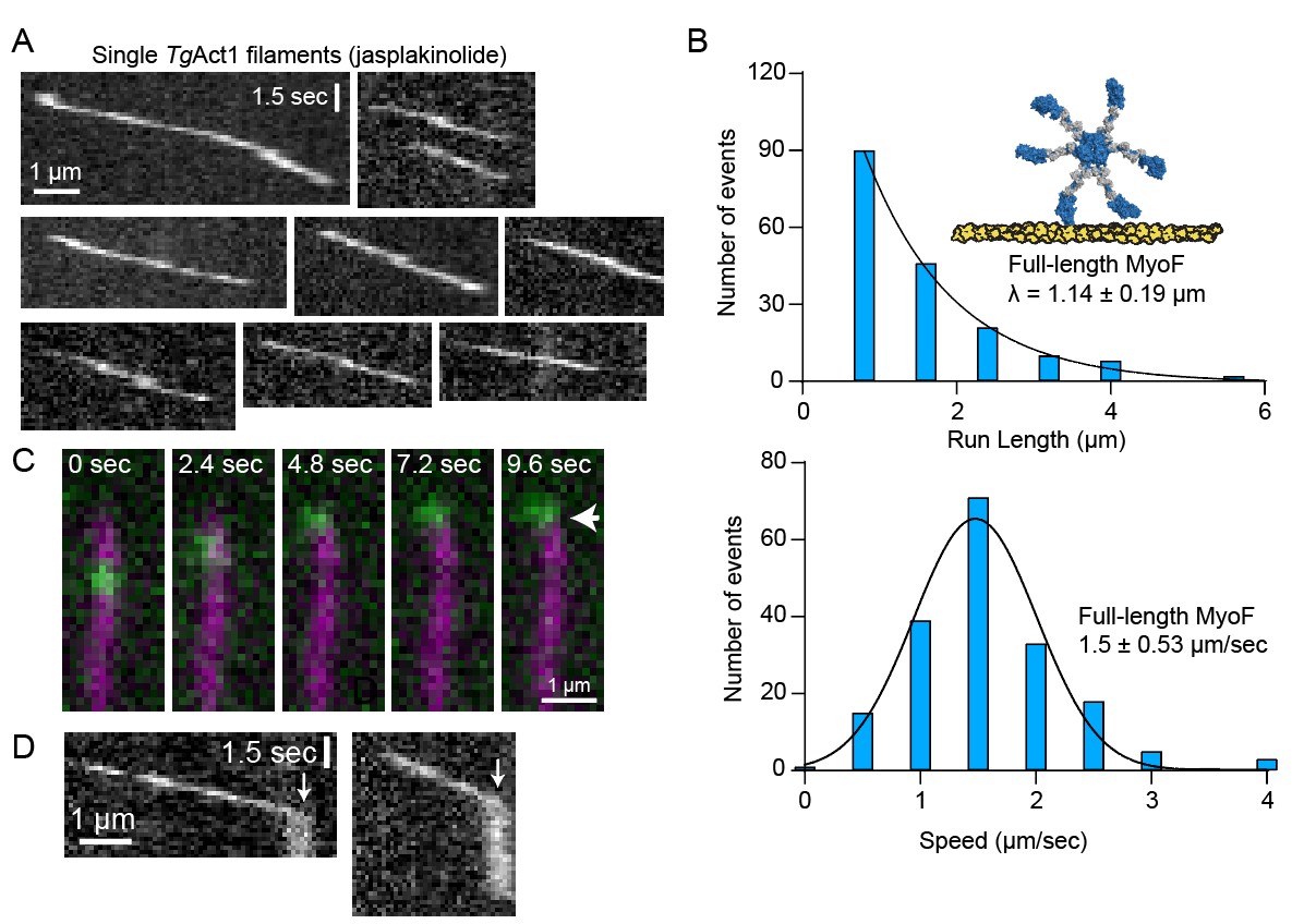
Toxoplasma gondii (T. gondii) is a single-celled Apicomplexan parasite that relies on a highly polarized endomembrane system for its invasion into and survival within host cells. Recent advancements in imaging technologies have revealed that vesicle transport and organization of organelles in the endomembrane pathway requires a highly dynamic actin cytoskeleton. These dynamics in turn rely on the activity of Myosin F (MyoF), a molecular motor unique to Alveolates. The defining characteristic of this molecular motor is a WD40 beta-propeller domain, exclusively found in this class of myosin. To understand the mechanism by which MyoF controls the dynamics and organization of actin, we studied the biophysical properties of the purified motor in vitro. A MyoF construct lacking its WD40 tail domain (MyoFΔtail) is dimeric and can bind and translocate actin in an in vitro motility assay. Single molecule studies show that the dimeric construct is non-processive however small ensembles move inefficiently on single filaments of skeletal actin. In contrast, single molecules of the full-length motor move processively on Toxoplasma actin and jasplakinolide-stabilized skeletal actin bundles. Electron microscopy of negatively stained images of MyoF and quantitative size exclusion chromatography shows that the WD40 domain oligomerizes to form a complex containing multiple dimeric molecules, which provides an explanation for why the full-length motor is processive compared to the dimeric MyoFΔtail construct. Finally, we show that MyoF binds microtubules through its WD40 domain and can slide actin filaments relative to microtubules. We propose a model whereby MyoF oligomers drive actin dynamics by translocating filaments relative to the parasite's cytoskeleton. These molecule features provide new insight into how MyoF functions in the cell to regulate actin organization during vesicle transport.
Bending stiffness of Toxoplasma gondii actin filaments
Wenxiang Cao, Thomas Sladewski, Aoife Heaslip, Enrique De La Cruz
Published in Journal of Biological Chemistry on December 18, 2024
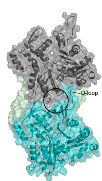
Actin is essential for the survival and pathogenicity of the Apicomplexan parasite Toxoplasma gondii, where it plays essential functions in cargo transport, invasion, egress, and organelle inheritance. Recent work has shown that, unlike vertebrate skeletal muscle actin, purified T. gondii actin filaments (TgAct1) can undergo rapid treadmilling, due to large differences in the barbed- and pointed-end critical concentrations, rapid subunit dissociation from filament ends, and a rapid nucleotide exchange rate constant from free monomers. Previous structural analysis suggested that the unique assembly properties of TgAct1 filaments may be a functional consequence of reduced contacts between the DNAse-1-binding loop (D-loop) of a filament subunit and its adjacent, long-axis subunit neighbor. Because the D-loop makes stabilizing interactions between neighboring subunits, it has been implicated in regulating the mechanical properties of actin filaments. In this study, we measured the bending persistence length (LB) of TgAct1 filaments and the filament length distribution. We found that despite compromised intersubunit D-loop contacts, TgAct1 filaments have similar bending stiffness and thermodynamic stability as vertebrate actin filaments. Analysis of published cryo-EM image density maps indicates that TgAct1 filaments retain a stabilizing intersubunit salt bridge between E168 and K62 and reveals visible density between Y167 and S61 of adjacent filament subunits, consistent with a conserved cation binding site proximal to the D-loop, as initially identified in vertebrate skeletal muscle actin filaments. These results favor a mechanism in which weak D-loop interactions compromise TgAct1 subunit incorporation at filament ends, while minimally affecting overall subunit interactions within filaments.
Toxoplasma gondii actin filaments are tuned for rapid disassembly and turnover
Kelli Hvorecny, Thomas Sladewski, Enrique De La Cruz, Justin Kollman & Aoife Heaslip
Published in Nature Communications on February 28, 2024
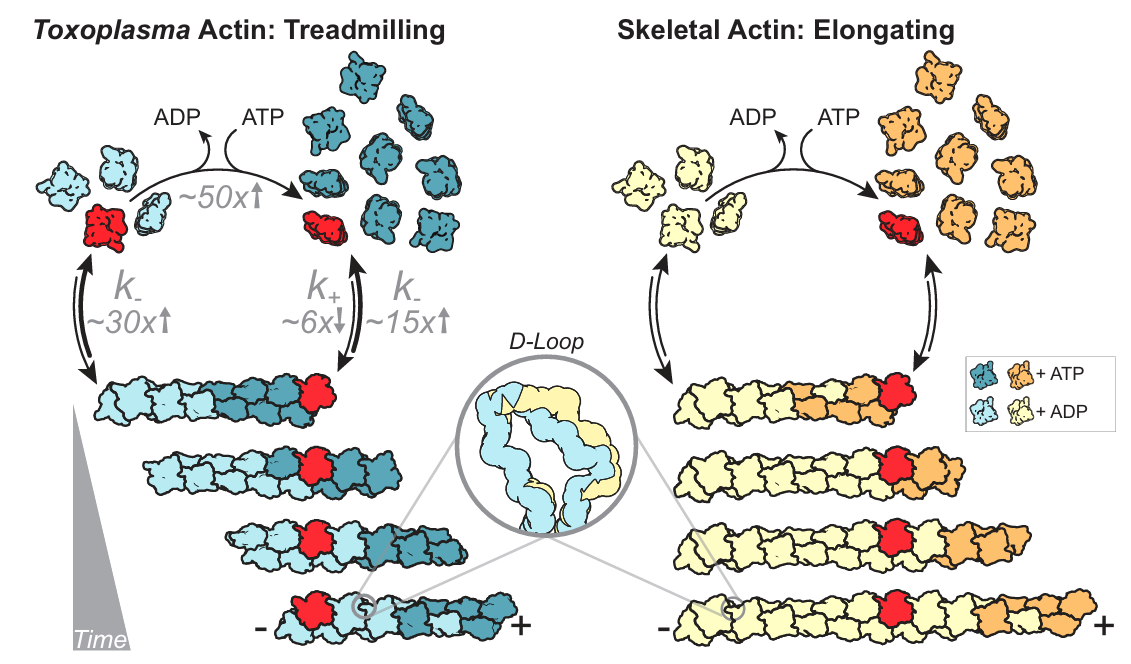
The cytoskeletal protein actin plays a critical role in the pathogenicity of the intracellular parasite, Toxoplasma gondii, mediating invasion and egress, cargo transport, and organelle inheritance. Advances in live cell imaging have revealed extensive filamentous actin networks in the Apicomplexan parasite, but there are conflicting data regarding the biochemical and biophysical properties of Toxoplasma actin. Here, we imaged the in vitro assembly of individual Toxoplasma actin filaments in real time, showing that native, unstabilized filaments grow tens of microns in length. Unlike skeletal muscle actin, Toxoplasma filaments intrinsically undergo rapid treadmilling due to a high critical concentration, fast monomer dissociation, and rapid nucleotide exchange. Cryo-EM structures of jasplakinolide-stabilized and native (i.e. unstabilized) filaments show an architecture like skeletal actin, with differences in assembly contacts in the D-loop that explain the dynamic nature of the filament, likely a conserved feature of Apicomplexan actin. This work demonstrates that evolutionary changes at assembly interfaces can tune the dynamic properties of actin filaments without disrupting their conserved structure.